


Valgus deformation of the feet is more often congenital. However, in some cases - with paralysis, traumatic injuries - they may already appear in the mature period of life. The main symptoms of pathology are pain in the area of the feet and leg muscles, a visible violation of the shape of the feet, as well as a change in march. The diagnosis of the disease is performed using a clinical examination, radius -x, electromyography, etc. Treatment includes conservative and surgical methods. However, proper efficacy is observed only in reconstructive operations.
What is this disease?
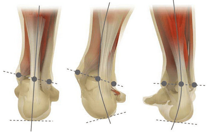
Valgus deformation is the curvature of the foot, characterized by the flattening of its longitudinal arc. Usually the inner edge of the foot is reduced ("drops") and the heel unfolds.
A person's foot, due to his location, assumes the pressure of the whole mass of the human body. For this reason, it has a special anatomical structure that allows depreciation, balance and stabilizing movements. However, an important component for the implementation of these tasks is the correct stop form.
Today, the most important problem of traumatology and orthopedics is the deformation of Valgus of the foot. The meeting is estimated at 30-58%, where 2/3 of cases make up congenital disorders.
Pathology is widely socially significant because it covers all age groups and also helps to curve the spine spine, the early development of osteochondrosis and arthrosis of the lower extremity joints.
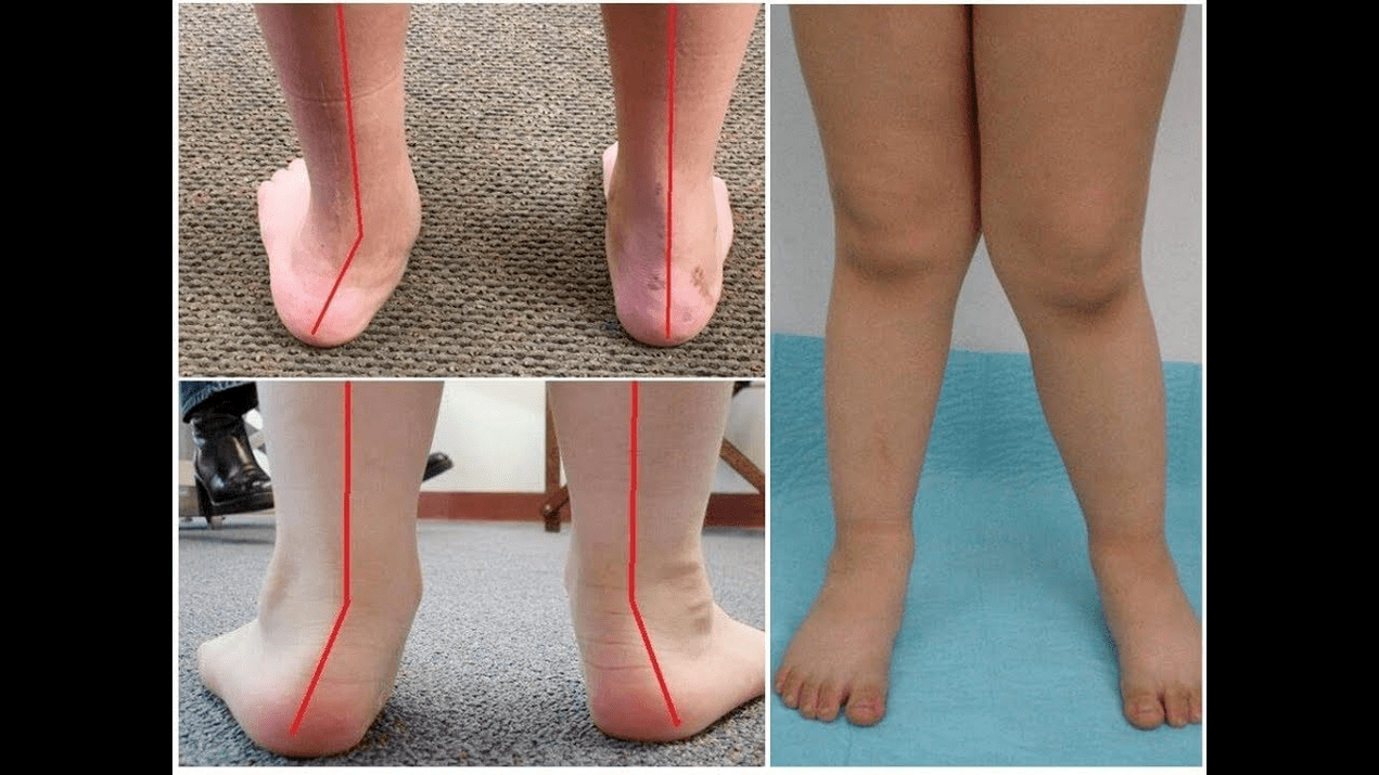
When you take your feet (if you look them from behind), a type X deformation is formed at the ankle level: the ankles are in contact, while the jumps are at a distance of 5 to 6 centimeters from each other.
Most of the time, the pathology is congenital in nature and is diagnosed in children back to the hospital (or immediately after the start of the walk). A similar condition is adjusted up to 5 years, after which (in the absence of proper treatment), a child develops flat feet.
Why does this arise?
It is believed that the main reason for the appearance of deformation of Valgus of the foot is an inadequate function of the posterior tibial muscle or weakness of the ligament apparatus.
Today, other factors in the development of pathology are distinguished:
- Congenital abnormalities with the incorrect location of the feet bones or the shortening of the tendons (vertical bone of the ram, short heap tendon);
- Posture disorders when the deformation of the feet compensates the curvature of the spine from the spine;
- Traumatic lesions (fractures of the bones of the feet, legs, hips or knees, ligament and tendon ruptures);
- Paralysis (immobilization) due to damage to the nervous system of encephalitis, polio, stroke, violation of cephalorean melting for hernias, etc. ;
- Spasm (constant contraction) of the leg muscles;
- Concomitant diseases: bone system pathology with vitamin D (rickets), diabetes mellitus, osteoporosis (reduction of bone density), impaired the thyroid and parathyroid glands, etc. ;
- Higher body weight, including a rapid weight gain in menopause or during pregnancy.
The development of pathology is also facilitated by incorrectly selected shoes or excessive correction of the club's foot in childhood.
Degree and stage of the disease
The severity of pathology (power of manifestation) is divided by degrees:
- Light with a 1, 5-2 centimeter vault height and a 15 degree heel tilt angle;
- The average, when the arc is flattened in the 1st century and the angle decreases to 10 degrees;
- Heavy at the height of the safe up to 0, 5 cm and the tilt angle of the heel is 5 degrees.
Depending on the involvement of certain structures, the following stages of curvature are distinguished:
- There are no bone deformations, the pain is determined on the inner surface of the ankle (in the posterior tibial muscle fixation area);
- The curvature is light, the heel is slightly rejected;
- The foot is allocated and the deformation is fixed (not correctly corrected);
- Weend is observed not only in the foot, but also in the ankle joint.
Symptoms
In the first step, patients are disturbed by periodic pain after prolonged walks or long vertical loads (standing or sitting on the foot). As a rule, pain syndrome intensifies when walking in incorrectly selected shoes. The next stage of the disease is associated with the occurrence of foot curvature: patients in the standing position do not depend on the outer edge of the foot, but with all their area. A slight change in march is observed.
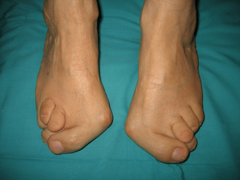
In the third stage, the protrusion of the thawed bone is determined (notably smaller than the ankle on the inner surface of the ankle), as well as a strong deviation from the outer heel (the patient is based on the inner edge of the heel bone). Advanced deformation of the feet valgus is characterized by a pronounced curvature of the foot itself and the ankle joint. Patients complain of intense pain in the leg muscles as well as a significant gear violation: the knees rub, while the right and left foot are located at some distance.
The severe curvature of the feet is often complicated by the deformation of the spine of the spine (scoliosis with different shoulder positions and pelvis wings), osteochondrosis (intervertebral disc damage with the formation of a hernia) or arthrosis (premature filling of intraarticular cartilage in the upper leg, round, the round.
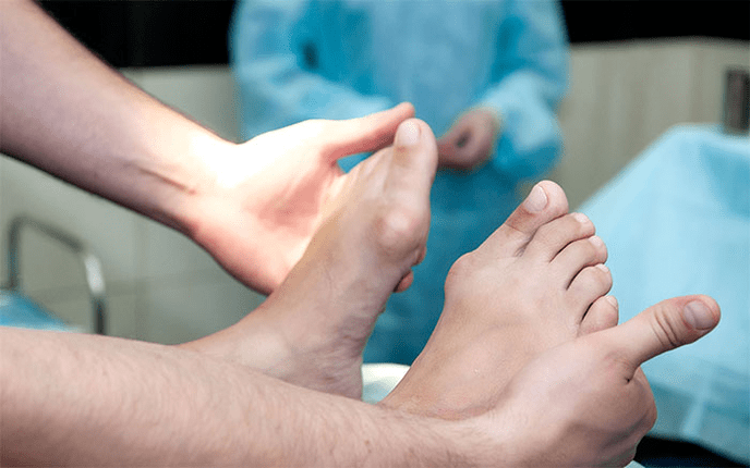
How to diagnose?
The diagnosis of the curvature of the foot consists of:
- Clinical inspection, during which the orthopedist detects a decrease in the arches of the foot, the diversion of the heel and the ram bones, the visible "disappearance" of the external and the protrusion of internal ankles.
- X -Ray - An accessible and informative method with which you can determine the change in the angles of the bone slope and linear parameters of your relationship. These indicators are necessary to make a final diagnosis and clarify the degree of deformation.
- The method of recording steps designed to determine the exact functional status of the limb. The method consists of recording the support time of individual parts of the foot when performing a step. Throughout the study, the phases of the roll of the foot are also studied, which reflect the balance of the lower limb muscles.
- Dynamic electromyography, which records the electrical activity of the studied muscles and their dependence on the step phase.
- Potoples amomography with digital processing that allows you to obtain all standard indicators and determine the type/degree of curvature with high precision.
An additional consultation of a neurologist (with deformations due to spasms or paralysis), endocrinologist (in the case of diabetes or thyroid/parathyroid gland disorders) and gynecologist (when the threatening) may be required. If the curvature of the foot appeared at the bottom of osteoporosis, densitometry is required - the study of bone density.
Treatment
Among the main methods of treatment of the curvature of Valgus from the feet, they are conservative and operational distinguished. Do not destroy painful joints with ointments and injections!
Conservative approach
This kind of help aims to get rid of the symptoms of the disease, but does not eliminate the root cause of the pathology.
The technique includes:
- the use of orthopedic insoles to support the I Plus bone, the foot arc, as well as the elimination of the middle and back of the foot lesions;
- Severe - Fixing the foot and ankle using special adhesive tapes with proper elasticity. Adhesive tape is used all the time within 3-5 days, after which are replaced;
- Sewing orthopedic shoes by individual patterns;
- The use of spelling and other foot and ankle fixation devices.
Conservative methods also include physiotherapeutic procedures (ozocnium, paraffin applications, electrophoresis, magnetic effects), massage and a complex of physiotherapy exercises, developed for a specific clinical case. Be careful! Today, most specialists prefer surgical treatment methods, because conservative therapy is ineffective (according to statistics, it is useless in 60% of cases).
Surgical intervention
The volume of operation and its type depend on the direct stage of the disease. Thus, the first degree of valgus deformation is treated by synovectomy (removal of the tendon shell for general voltage correction) or the heel osteotomy (dissection) to return to anatomically correct position. In the second stage of disease development, transplantation of fingertips curves is used. This intervention is generally performed against the heel dissection fund or a loba of Carneiro (surgical immobilization of the articulation between the bones of the RAM and the scaphoid).
The curvature of grade III requires arthrodesis of various joints of the foot at once: the freer, five cubic cubic and ram. This immobilization three times is often complemented by heel bone dissection. In phase IV of the pathology, reconstructive operations are necessary not only in the foot but also on the ankle. In this case, the instability of the ligament apparatus is adjusted using transplants (from its own body or artificial materials). The volume of operations on the foot itself is the same as the degree of curvature III.
Recovery period
Rehabilitation includes walking without support in the operated leg for 2 months. At the same time, the patient needs to use removable plaster longShit from 1, 5 to 3 months. Active movements in the operated foot are recommended to start after 1, 5 months after surgery. In the third month, a physical education strengthening complex is introduced. However, later, patients are prohibited from accidents and active sports activities. It is worth noting that it is possible to judge the final result of the surgery only six months later.
Preventive measures
The prevention of the deformation of Valgus of the Parade includes the following measures:
- Early correction of congenital anomalies with the inadequate arrangement of the feet bones or shortening of the tendon grains (vertical thawing bone, short jump tendon);
- Correction of posture disorders (scoliosis, etc. );
- Timely treatment of traumatic lesions (fractures of the feet, leg, thigh or knee joint, ligament and tendon device);
- Correct rehabilitation after paralysis (immobilization) due to damage to the nervous system for encephalitis, polio, stroke, cephalraquidal root violation for hernias, etc. ;
- Spasm relief (constant reduction) of the leg muscles;
- Therapy for concomitant diseases: bone system pathologies with vitamin D (rickets), diabetes mellitus, osteoporosis (reduction of bone density), impaired function of thyroid and paratyroid glands, etc. ;
- A decrease in normal body weight (especially with rapid post -feedback weight gain or due to pregnancy);
- Selection of orthopedic shoes or the use of supinators;
- Moderate Foot Correction of the No Treatment Club of Hyper-Correction-Treatment that leads to secondary excretion of Valgo from the feet.
Prevention of disease progression is to use conservative methods and early reconstructive operations. In this case, physical activity is limited to prevent destruction and curvature of the ankle joints. Remember that the timely treatment of the deformation of Valgus from the feet not only improves the quality of life of patients, but also prevents the development of osteochondrosis and arthrosis of knee or hip joints!